
CT Technique and Accuracy
| Value of rectal contrast administration for
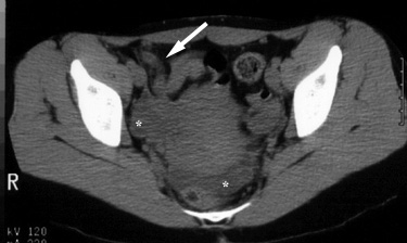
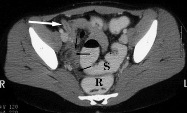
CT. A. The left-hand image below was obtained without intravenous, oral or rectal contrast administration. It demonstrates an inflamed appendix (arrow). Note fluid in the right adnexa and cul-de-sac regions (*). Differentiation between fluid-filled bowel loops, free fluid, and abscess is difficult. B. The right-hand image below was obtained after administration of intravenous and rectal contrast. Differentiation of the rectosigmoid colon (R, S) from abnormal fluid collections is facilitated. Abnormal fluid is identified in the right pelvis (black arrows). The inflamed appendix with enhancing walls (white arrows) is also better delineated. |
 |
 |
Return to CT Technique and Accuracy