CT
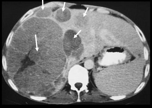
Multicentric hepatoma |
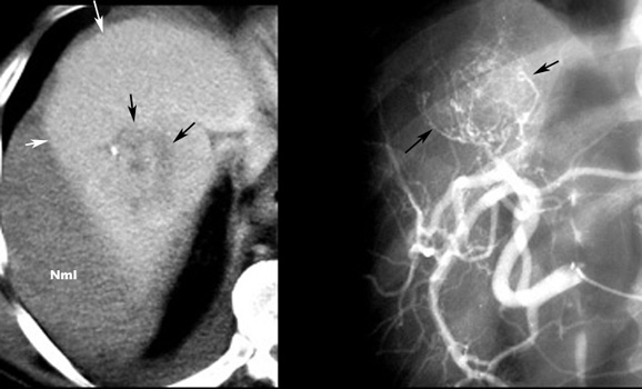 |
Hepatoma with hemorrhage
|
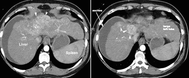 |
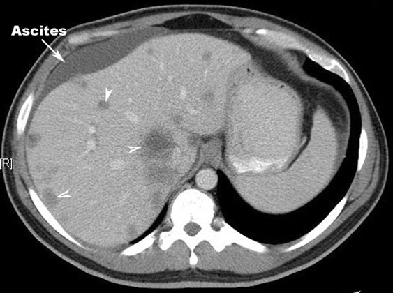
HepatomaArrowheads point to the enhancing mass. Note the lobulated margins of the liver. Liver has lower density than spleen and ascites indicating underlying cirrhosis. |
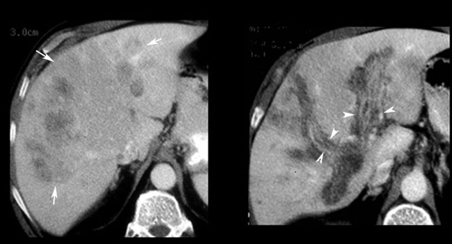 |
Hepatoma
|
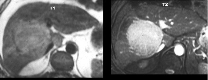 |
HepatomaMR shows a mass that has low signal intensity on T1 and high signal on T2. |
 |
Liver metastasisMultiple hypodense lesions seen in the liver with no significant contrast enhancement. Primary: Colon carcinoma |