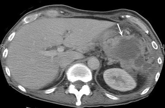
Subdiaphragmatic abscess post splenectomy
- Arrow points to multiloculated thick walled fluid collection in the left upper quadrant of the abdomen.
- Note absence of spleen.
Sub diaphragmatic
| |
Subdiaphragmatic abscess post splenectomy
|
Pancreatic
 |
Pancreatic abscessDiffusely enlarged pancreas with air pockets. Arrow points to body of Pancreas Abscess is in tail of Pancreas |
Renal
|
|
Renal abscess
|
|
Renal abscessCT shows a large mass in the left renal area with multiple air pockets and absence of functioning renal parenchyma. |
Appendiceal
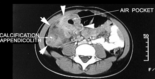 |
Acute Appendicitis Appendiceal abscessCT Post Contrast:
|
Liver
Diverticulitis
Post Diverticular abscess drainage |


