Principle
How it is done
- X-rays are high energy radiation waves that can penetrate body parts to produce an image.
- Patient is positioned so that the body part to be evaluated is between the x-ray source and a device (cassette) that records the image.
- The patient must remain still (for a couple seconds) when the x-ray is taken.
- Most plain x-ray examinations consist of at least two views of a body part at right angles to one another (i.e. PA and lateral CXR).
Example Indications
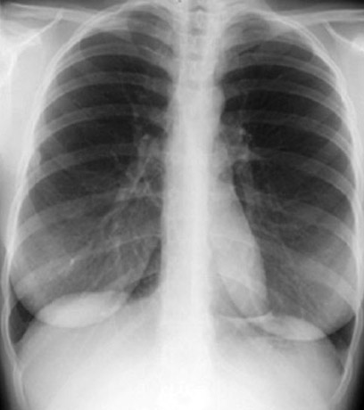
- Chest x-ray
- Most commonly used imaging procedure
- Practically done in most hospitalized patients
- For evaluation of pulmonary or cardiac symptoms
- Abdomen
- Acute pain
- Bones and Joints
- Injuries
- Pain
Limitation
- Significant pathology can be missed
- Radiation exposure
Useful for
- It can be diagnostic with no additional imaging required.
- It serves as preliminary image to plan for more specific imaging studies like CT or MRI.
- Required for interpretation of other imaging procedures like V/Q lung scan.
Cost: $
Densities
Air: Dark
Liquid density: White
Fat: Gray
Bone: Dense white
Metal: Extremely dense white