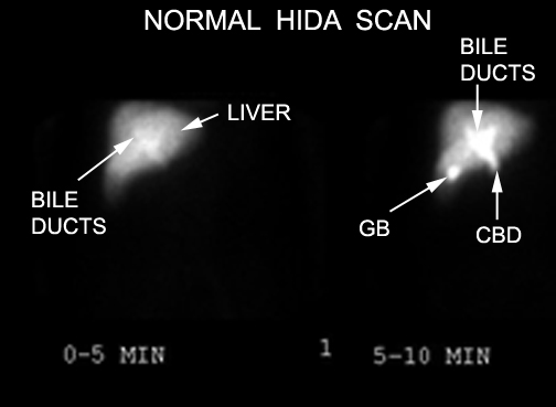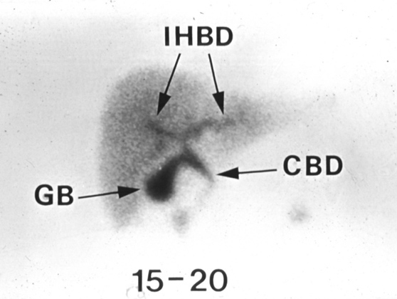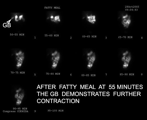
HIDA Scan
Normal scan After fatty meal
A HIDA scan is an imaging test used to examine the gallbladder and the ducts leading into and out of the gallbladder.
How it is done
- The patient receives an intravenous injection of a radioactive material called hydroxy iminodiacetic acid (HIDA).
- The HIDA material is taken up by the liver and excreted into the biliary tract.
- In a healthy person, HIDA will pass through the bile ducts and into the cystic duct to enter the gallbladder.
- It will also pass into the common bile duct and enter the small intestine, from which it eventually makes its way out of the body in the stool.
- HIDA imaging is done by a nuclear scanner, which takes pictures of the patient's biliary tract over the course of about two hours.
- The images are then examined by a radiologist, who interprets the results.
Safety
- It is generally a very safe test and is well tolerated by most patients.
Indication
- The HIDA scan should be done when ultrasound is not diagnostic and when there is a clinical diagnosis of acute cholecystitis.
- Usually, HIDA scans are ordered for patients who are suspected of having an obstruction in the biliary tract, most commonly those who are thought to have a stone blocking the cystic duct leading out of the gallbladder.
- Such a scenario is consistent with acute cholecystitis, which often requires surgical removal of the gallbladder.
- In cholecystitis, HIDA will appear in the bile ducts, but it will not enter the cystic duct or the gallbladder -- a finding that indicates obstruction.
- If the HIDA enters the bile ducts but does not enter the small intestine, then an obstruction of the bile duct (usually due to stones or cancer) is suspected.
Limitation
- HIDA scans can be falsely positive when the gallbladder is not filling in the absence of cholecystitis. These situations include severe liver disease, patients on total parenteral nutrition, hyperbilirubinemia, inadequate fasting, and alcohol and opiate abuse.
- In the presence of cystic duct obstruction the gall bladder does not fill with isotope.
Interpretation
A normal result means that the gallbladder is visualized within 1 hour of the injection and the tracer is in the small intestine.
GB not visualized: If the gallbladder is not visualized within 4 hours after the injection it indicates that there is either cholecystitis or cystic duct obstruction.
HIDA scan for acute cholecystitis has a sensitivity of 97%, Specificity of 94%.
Tracer not visualized in intestines means common bile duct obstruction. If the radioactive tracer moves through bile ducts very slowly, this may indicate a blockage or obstruction. Or it may indicate a problem in liver. .
If the radioactive tracer is found outside of biliary system it indicates a leak.
Cost
- $$

