 |
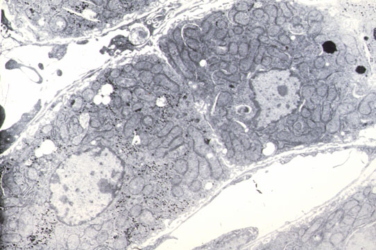
They drain bile from the hepatocytes to the portal space. Microscopically visible only when distended by bile. Can also be demonstrated by Gomori's reaction for alkaline phosphatase and silver impregnation. Their wall is made by the plasma membrane of two adjacent hepatocytes which form intracanalicular villi. They form an intricate anastomotic network of canals.
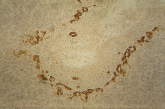 |
In the periportal region the biliary tree takes origin. The bile canaliculus lined by hepatocytes meets the ductule, first lined by hepatocytes and ductal epithelium, then lined only by ductal cells. They penetrate the portal space and connect with the interlobular bile duct. Bile ductules have a basal lamina. The ductal epithelium is cuboidal with light stained cytoplasm, dark nuclei and few cytoplasmic organelles.